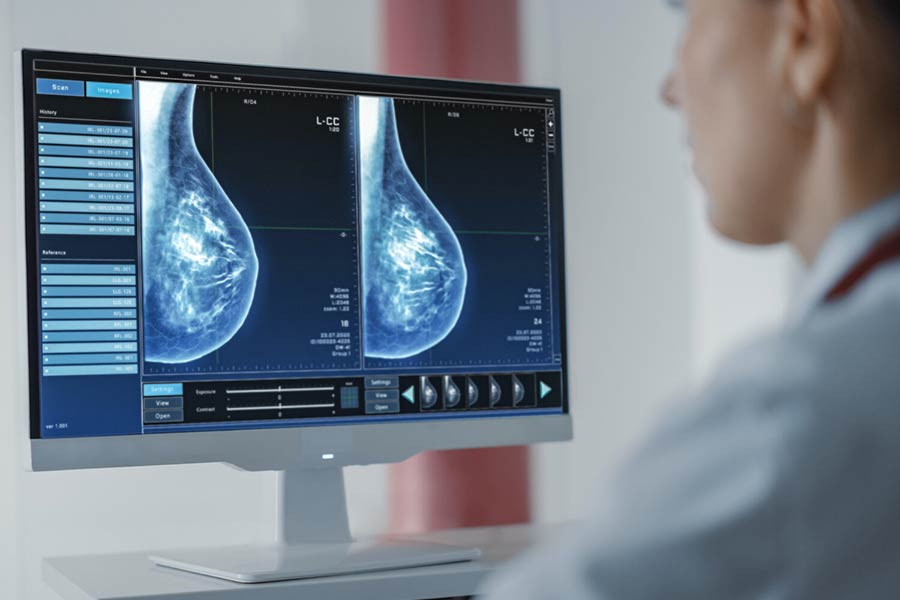
Sadly, around one in eight (13%) of American women will develop invasive breast cancer over the course of their lifetime, with 2021 figures estimating that there will be around 281,550 new cases of invasive breast cancer this year in America alone.
Approximately 2.1 million women were diagnosed globally with breast cancer in 2018, of which 600, 000 did not survive. Breast cancer remains the most common form of cancer in women and many of us, heartbreakingly, will be touched by the disease during our lifetimes, whether through the experience of seeing a loved one, colleague or neighbor cope with the disease, or even by receiving a diagnosis ourselves.
Mammograms already offer a gold standard in terms of the diagnosis of breast cancer. However, mammography carries its limitations, particularly when it comes to early breast cancer diagnosis. Mammography can generally only detect breast cancer when the disease has already progressed significantly to the point that a mass, or tumor, can be detected, and currently, only women over the age of 50 are offered routine mammogram scans, removing the potential for early diagnosis of the disease from the under 50 age-group entirely. Despite this age restriction, around 11% of breast cancers occur in women under the age of 45, with an estimated 26, 393 women under 45 expected to be diagnosed in the US this year. Studies now indicate that mammograms also generally fail to detect around 20-30% of breast cancer tumors even in the later stages of development.
Whilst mammography is still considered the gold standard for breast cancer diagnosis, the NIHR (National Institute for Health Research) nevertheless reports that early detection via screening women for breast cancer in their 40’s would reduce their chance of dying from breast cancer by 25% in the first ten years of screening. The general consensus amongst clinicians is that the most powerful weapon we have in the fight against breast cancer mortality is early detection, yet the efficiency of mammograms drops as low as 50% in detecting cancer in women with dense breast tissue, and mammograms can only detect cancer when the disease has progressed to the formation of a small tumor or lesion.
Similarly, a recent 23-year longitudinal study published in The Lancet Oncology reported 11.5 years of life saved per 1,000 women who received screening in their forties – yet how many women under fifty receive it? The answer is, very few unless there is a pre-existing medical rationale for referring the patient for screening. As Stephen Duffy, Professor of Cancer Screening at Queen Mary University of London commented so sagely, ‘Breast cancer does not respect one’s 50th birthday.’
Tantalizingly, a form of screening known as thermography offers an early adjunct to mammography as a potential means of breast cancer detection, offering the potential to detect cancer lesion development in its earliest stages in a non-invasive and pain-free way. The potential to catch the disease at an early, asymptomatic stage carries great promise in canceling out the need for more aggressive, later-stage treatment (such as chemotherapy) where cancer has grown or metastasized.
A specific kind of thermography known as DITI (Digital Infrared Thermal Imaging) has already been approved by the FDA and offers a promising and effective adjunct treatment to mammography, in that it offers a risk-free, pain-free, non-invasive, and radiation-free means of identifying anomalies in breast tissue temperature, metabolism and blood flow, an activity that could be indicative of breast cancer. It is also relatively portable, cost-effective, and accessible to women of all ages.
Let’s consider the merits of this fascinating adjunct screening technology and consider how it might be of benefit to you or your family.
Early Detection with Breast Thermography
Breast thermography (DITI) works by using an extremely sensitive heat camera that produces high-resolution, infrared photographs of the breast. Extremely subtle changes in temperature and blood flow can be closely monitored over time simply by observing changes in temperature, with comparative data of each breast compared for adaptations and anomalies, and ‘hot spots’ potentially indicating a cancerous growth.
DITI is not a diagnostic tool, in the sense that shifts in heat patterns and vascular activity might be attributable to a whole range of factors, many non-cancerous (e.g., a benign growth, infections, fibrocystic disease, lymphatic activity, hormonal dysfunction, inflammation, or vascular disease) – so the screening must be carried out by a competent physician or clinician who possesses the adequate knowledge of the patients’ history and the technical competency to evaluate results and recommend and explore further treatment options.
The reason that adaptations in localised body temperature and blood flow can be so illuminating in the early detection of cancerous tumors is that cancer cells usually exhibit the need for an increased blood supply (i.e., angiogenesis). Since tumors divide and grow rapidly, this will also likely lead to an increased localized metabolic rate over time, with DITI allowing close monitoring of upward shifts and temperature gradients that might be indicative of a lesion or tumor growing in that area of the breast.
DITI offers an added potential benefit, in that it makes screening more accessible to wider populations: a recent pilot study in India concluded that thermal imaging techniques offered a positive means of reaching a large sector of the population to whom mammographic scanning remained generally inaccessible (whether for age-related, or geographic reasons). The study involved 1, 008 female patients aged 20-60 years of age, who had not received a diagnosis of breast cancer, reporting that of the 49 subjects who exhibited abnormal results, 41 were found via the subsequent use of clinical, radiological, and histopathological treatments to have breast cancer.
The remaining 8 subjects in that abnormal group were found to have exhibited anomalous results either because they were breastfeeding, or because of fibrocystic breast disease. The DITI study reported a sensitivity of 97.6% as a screening modality, with the specificity of 99.17%, and a positive predictive rate of 83.67%, with researchers concluding that thermal imaging scans offered a highly effective adjunct to early cancer diagnoses.
Click here to know more about Thermography.
Considerations
Thermographic scan accuracy and efficacy have been observed to be mediated and influenced by a range of factors, such as the utilization of exogenous steroid therapy by the patient, pregnancy, lactation, the onset of menopause, and during specific stages of the menstrual cycle stage. The thermographic examination must be undertaken by qualified practitioners in a draft-free, temperature and humidity-controlled room with a constant temperature of 20 degrees.
Summary
Breast cancer can originate in any part of the breast, beginning either in milk-carrying ducts or in the milk-producing lobules of the breast. As cancer cells generate heat, due to the release of nitric oxide into the blood, this leads to an observable alteration of microcirculation rates.
When cancer cells proliferate, vasodilation takes place in order that blood circulation increases, and neo-angiogenesis (the creation of new blood vessels to supply nutrients to the tumor) can occur, with the metabolic activity of cancerous cells increasing concomitantly. By efficiently and quickly identifying these localized physiological changes via thermal imaging techniques, DITI offers an excellent potential means of detecting breast cancer and its earliest stages.
As breast cancer rates continue to increase on a global level, innovations in early detection remain a key weapon, not only in the effective prevention and management of the disease, but also in the lowering of mortality rates, and in the prevention of the need for more aggressive cancer treatment interventions as a result of late-stage diagnosis.
References
National Institute for Health Research (NIHR). (2020). Case Study: Breast Cancer Screening for Women in their Forties Could Save Lives. Accessible at: https://www.nihr.ac.uk/case-studies/breast-cancer-screening-for-women-in-their-forties-could-save-lives/26328
Omranipour, R., Kazemian, A., Alipour, S., et al. (2016). Comparison of the Accuracy of Thermography and Mammography in the Detection of Breast Cancer. Breast Care (Basel). 11, 4, pp. 260-264.
Rassiwala, M., Mathur, P., Mathur, R., Farid, K., Shukla, S., Gupta, P.K., Jain, B. (2014). Evaluation of digital infra-red thermal imaging as an adjunctive screening method for breast carcinoma: a pilot study. International Journal of Surgery. 12, 12. Pp.1439-43.
Singh, D. Singh, A.K. (2020). Role of image thermography in early breast cancer detection- Past, present and future. Computer Methods and Programs in Biomedicine, 183, 105074. Accessible at: https://www.sciencedirect.com/science/article/abs/pii/S0169260719311277
___________________________________________
These statements have not been evaluated by the Food and Drug Administration. This product is not intended to diagnose, treat, cure or prevent any disease.
Use only as directed. Consult your healthcare provider before using supplements or providing supplements to children under the age of 18. The information provided herein is intended for your general knowledge only and is not intended to be, nor is it, medical advice or a substitute for medical advice. If you have or suspect you have a specific medical condition or disease, please consult your healthcare provider.
First Published: © 2021 Doctors Studios

